Patellar Instability and Dislocations
What Is Patellar Instability?
Patellar instability refers to a condition where the kneecap (patella) moves out of its normal groove on the thigh bone (femur), either partially (subluxation) or completely (dislocation). This condition may be traumatic (from injury) or chronic/recurrent due to underlying anatomical issues.
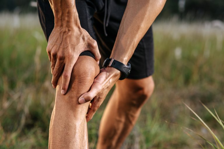
It most commonly affects young athletes, particularly females, and those involved in sports that require cutting, twisting, or jumping motions.
Causes and Risk Factors
Several factors can contribute to patellar instability or dislocation:
Direct trauma to the knee (common in sports or falls)
Loose or stretched ligaments (especially the MPFL — medial patellofemoral ligament)
Shallow trochlear groove (where the kneecap normally glides)
High-riding kneecap (patella alta)
Knock-knee alignment (valgus knee)
Generalized ligament laxity (hypermobile joints)
Symptoms of Patellar Dislocation
Sudden pain and deformity after a twisting injury or direct blow
Visible displacement of the kneecap to the outside of the leg
Inability to straighten or bear weight on the knee
Swelling and bruising after the event
A feeling of looseness or slipping of the kneecap during activity
Diagnosis
Physical exam: Your provider will check for ligament damage, kneecap mobility, and alignment.
X-rays: Assess bone structure, patella positioning, and possible fractures.
MRI: Evaluates cartilage, ligament injury (especially MPFL), and other soft tissue damage.
Treatment Options
Non-Surgical Management (for first-time dislocations)
Manual reduction: Putting the kneecap back into position
Knee brace or immobilizer: Used short-term to protect healing
Physical therapy:
Strengthens quadriceps, especially the vastus medialis obliquus (VMO)
Corrects alignment and movement patterns
Activity modification to prevent recurrence
Surgical Treatment (often needed for recurrent instability)
MPFL reconstruction: Rebuilding the ligament that holds the patella in place
Tibial tubercle osteotomy (TTO): Repositioning the tendon attachment for better alignment
Trochleoplasty: Reshaping the groove where the kneecap sits (rare and complex)
Lateral release: Releasing tight tissues on the outer knee (used selectively)
Recovery and Return to Sport
Non-surgical: 4–8 weeks with proper therapy
Surgical: 4–6 months depending on the procedure
Return-to-play testing is recommended for athletes
A personalized rehab plan, created by your team at Kerlan Jobe Institute, is crucial for restoring stability and preventing future episodes.
When to Consider Surgery
Surgery may be appropriate if you:
Have had multiple dislocations
Have persistent pain or instability despite rehab
Have anatomical abnormalities contributing to patellar tracking issues
Are a competitive athlete aiming to return to sport safely
Patellar Stability Is Possible
With expert evaluation and a personalized treatment plan, most patients return to full function — and even high-level athletics — after addressing patellar instability. The orthopedic team at Kerlan Jobe Institute specializes in knee realignment procedures, ligament reconstruction, and recovery plans built around each patient’s lifestyle and goals.