Cartilage and Bone Injuries Within the Ankle Joint
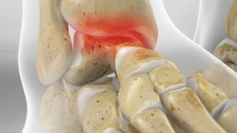
What Are Osteochondral Lesions of the Talus (OLT)?
Osteochondral lesions of the talus (OLT) are injuries that involve both the cartilage and the underlying bone of the talus, one of the main bones in the ankle joint. These lesions can result from a single traumatic event such as an ankle sprain or fracture, or from repetitive microtrauma over time. OLTs may also be referred to as osteochondritis dissecans or talar dome lesions and can lead to chronic pain, swelling, and mechanical instability of the ankle.
Causes and Risk Factors
- Acute ankle injuries, particularly severe sprains or fractures
- Repetitive ankle trauma (e.g., in athletes or dancers)
- Poor healing from previous ankle injuries
- Congenital alignment abnormalities
- Limited blood supply to the talar dome (contributing to poor cartilage healing)
Symptoms
- Deep ankle pain, often localized to the inner or outer side
- Swelling that may worsen with activity
- Ankle stiffness and limited motion
- Locking or catching sensations
- A feeling of instability or “giving way”
- Persistent symptoms despite previous ankle sprain treatment
Diagnosis
- Physical exam may reveal joint tenderness or limited motion
- X-rays can show bone abnormalities but may miss cartilage injuries
- MRI provides detailed imaging of cartilage damage and bone edema
- CT scan may be used for surgical planning in advanced or fragmented lesions
Treatment
Non-Surgical Treatment
- Immobilization and reduced weight-bearing (typically 6–8 weeks)
- Activity modification and anti-inflammatory medications
- Physical therapy to restore range of motion and strength
- Ankle bracing or supportive orthotics
- For small or stable lesions, conservative management may provide relief
Surgical Treatment
- Arthroscopic debridement and microfracture: Stimulates new cartilage growth in smaller, contained lesions
- Osteochondral autograft or allograft transplantation: Replaces damaged cartilage and bone with healthy tissue
- Retrograde drilling: Enhances healing by creating channels to stimulate blood flow
- Fixation of loose fragments: When a bone/cartilage fragment is intact and viable
- Autologous chondrocyte implantation (ACI): A specialized technique used in select cases to regenerate cartilage
Recovery Timeline
- Non-surgical recovery: 6–12 weeks of immobilization and rehab
- Surgical recovery:
- Weightbearing restrictions for 6–8 weeks
- Physical therapy begins during early healing
- Gradual return to sports and high-impact activities over 4–6 months
- Full recovery for cartilage transplantation or grafting: 6–12 months
- Full return to activity depends on procedure type and individual progress
Expert Treatment at Kerlan Jobe Institute
The specialists at Kerlan Jobe Institute are experienced in diagnosing and treating OLTs with precision and care. Whether using conservative management or advanced surgical techniques, our team prioritizes joint preservation, long-term mobility, and return to activity.