Chondromalacia Patella
Softening of the Cartilage Behind the Kneecap
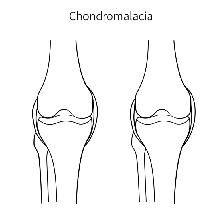
What Is Chondromalacia Patella?
Chondromalacia Patella refers to the softening and breakdown of the articular cartilage on the undersurface of the kneecap (patella). This cartilage normally allows the kneecap to glide smoothly over the femur. When it softens or deteriorates, it can cause pain, inflammation, and grinding sensations during knee movement.
While it’s sometimes used interchangeably with Patellofemoral Pain Syndrome (PFPS), chondromalacia specifically refers to structural cartilage damage, whereas PFPS may not.
Who Is at Risk?
Chondromalacia Patella is especially common among:
Adolescents and young adults
Runners, cyclists, and athletes in high-impact or repetitive sports
People with previous patellar dislocation or instability
Individuals with muscle imbalances, malalignment, or flat feet
Symptoms
Pain at the front of the knee, especially behind the kneecap
Worsening pain with stairs, squatting, kneeling, or prolonged sitting
Grinding or cracking sensations (crepitus) when bending the knee
Occasional swelling after activity
Stiffness or difficulty fully straightening the leg
Causes and Contributing Factors
Chondromalacia can be triggered or worsened by:
Poor patellar tracking due to muscle imbalance or alignment issues
Overuse from repetitive motion (e.g., running, jumping)
Kneecap trauma, such as falls or direct impact
Weak hip or thigh muscles
Tight hamstrings or IT band
Diagnosis
Diagnosis begins with a physical exam focused on:
Patellar mobility
Quadriceps strength
Signs of malalignment or tracking issues
Imaging such as X-rays or MRI may be used to:
Assess cartilage quality
Identify patellar positioning issues
Rule out other causes of knee pain
Treatment Options
Non-Surgical Treatment
Most patients respond well to conservative care:
Physical therapy to:
Strengthen the quadriceps and hip stabilizers
Improve patellar tracking
Increase flexibility in the hamstrings and IT band
Ice and NSAIDs to reduce inflammation
Activity modification: Avoid deep squatting, stairs, or prolonged sitting
Orthotics or taping for alignment correction
Patellar bracing to improve tracking
Surgical Options (For severe or non-responsive cases)
Arthroscopy to remove damaged cartilage or smooth surfaces
Lateral release if tight lateral structures are pulling the patella out of alignment
Cartilage restoration procedures (in select cases)
Recovery and Prognosis
Most patients improve in 6–12 weeks with proper rehabilitation
Long-term results are best with consistent strengthening and alignment correction
Return to sports is possible but may require modification in training habits
Kerlan Jobe Institute’s Approach
At Kerlan Jobe Institute, we recognize the importance of individualized care. Our specialists conduct a thorough biomechanical assessment to target the root cause — not just the symptoms — of patellofemoral cartilage pain.