Understanding Knee Anatomy
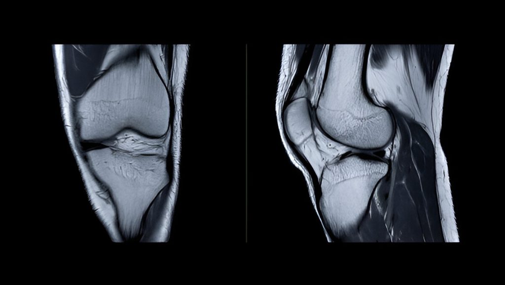
The knee is one of the most powerful and intricate joints in the human body. It supports your weight, absorbs shock, and enables a wide range of motion. Whether you’re a professional athlete or just climbing stairs, your knees are constantly at work.
The Bones of the Knee
Femur (thighbone) – Connects the hip to the knee.
Tibia (shinbone) – Supports the body’s weight below the knee.
Patella (kneecap) – Protects the front of the knee and improves muscle efficiency.
These bones articulate at the tibiofemoral and patellofemoral joints.
Ligaments: The Knee’s Stabilizers
The knee is stabilized by four key ligaments:
ACL (Anterior Cruciate Ligament) – Controls rotation and forward motion of the tibia.
PCL (Posterior Cruciate Ligament) – Controls backward motion of the tibia.
MCL (Medial Collateral Ligament) – Stabilizes the inner knee.
LCL (Lateral Collateral Ligament) – Stabilizes the outer knee.

Cartilage: The Knee’s Cushion
Two types of cartilage provide support and shock absorption:
Articular cartilage – Smooth tissue covering bone ends to reduce friction.
Meniscus (Medial & Lateral) – C-shaped fibrocartilage acting as shock absorbers and stabilizers.
Muscles and Tendons: Power and Control
Major muscle groups driving knee movement include:
Quadriceps – Straighten the knee.
Hamstrings – Bend the knee.
Gastrocnemius & Soleus – Support motion from the calf.
Patellar Tendon – Connects the quadriceps to the tibia through the kneecap.
Synovial Membrane and Bursae
The synovial membrane lines the joint and produces synovial fluid for lubrication.
Bursae are small fluid-filled sacs that prevent friction between bones, tendons, and skin.
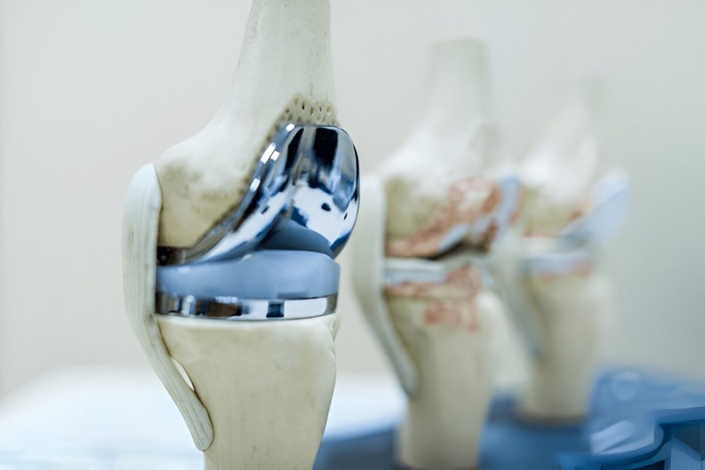
Why It Matters
Knowledge of knee anatomy helps:
Diagnose and treat injuries like ACL tears, meniscus damage, and arthritis
Design better rehab and performance plans
Prevent long-term damage through early intervention