Dr. Jung provides tips and tactics.
Article: Ottawa Ankle Rules – Ankle Xray Criteria for Adults
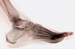
The Ottawa Ankle Rule screens for fractures of the ankle as well as the mid-foot region. Research suggests that a patient presenting with none of the Ankle Rule symptoms is less than 1% likely to have a fracture.
Ankle injuries are a significant medical and socioeconomic problem, and traumatic injuries to the ankle are common. However, unnecessary X-rays can expose patients to radiation and affect their risk of developing cancer, while also increasing medical costs, according to the Food and Drug Administration.
The Ottawa Ankle Rule utilizes information on bony tenderness (at specific landmarks, particularly the malleoli), as well as patient ability to bear weight to determine if an x-ray is advisable.
An ankle x-ray series is indicated if there is any pain in the malleolar zones and any one of the following:
-
bone tenderness along the distal 6 cm of the posterior edge of the tibia or tip of the medial malleolus, or
-
bone tenderness along the distal 6 cm of the posterior edge of the fibula or tip of the lateral malleolus, or
-
inability to bear weight both immediately and in the emergency department for four steps
A foot X-ray series is indicated if there is any pain in the midfoot zone and any one of the following:
-
bone tenderness at the base of the fifth metatarsal (for foot injuries), or
-
bone tenderness at the navicular bone (for foot injuries), or
-
inability to bear weight both immediately and in the emergency department for four steps
Tips & Tactics provided by Kenneth Jung, MD, board-certified orthopedic foot and ankle surgeon at Cedars-Sinai Kerlan-Jobe Institute in Los Angeles.
The Ottawa Ankle Rule was developed by emergency physicians to assess common musculoskeletal injuries and is useful in guiding clinical decision-making. As with any guide, clinical judgment plays a role as well. The clinical presentation of foot and ankle injuries may evolve with time; thus, initial presentation utilized by the Ottawa Ankle Rule may differ from the clinical presentation in the practitioner’s office. Swelling, tenderness, bruising, and ecchymosis will evolve to diffuse symptoms over the course of days which can alter the physical examination.
History of swelling is important. Initial swelling localized to soft tissue versus bone landmarks can be useful in identifying the injured structure(s). With abundant mobile devices present, patients often have pictures readily available for review. They may document the progression of symptoms of swelling, bruising, and ecchymosis. This information, along with the reported location of initial pain, may guide the physician as to whether the injured structure is bone or soft tissue.
In my clinical practice, I utilize the palpation component of clinical examination to localize symptoms – pain to bone and soft tissue structures. I specifically palpate the following bony landmarks: lateral malleolus, medial malleolus, anterior process of calcaneus, fifth metatarsal base, fifth metatarsal shaft, and navicular. Soft tissue landmarks include the deltoid ligament, posterior tibial tendon, lateral collateral ligaments, peroneal tendons (retromalleolar), peroneal tendons (distal to lateral malleolus), and anterior tibiotalar joint capsule. With the Ottawa Ankle Rule as a guideline, radiographs are indicated for localized bony tenderness.
Concomitant bone and soft-tissue injuries may also exist. Radiographs can be utilized to diagnose fractures as well as assess the bony alignment. Ligamentous injuries to the deltoid, syndesmotic, and Lisfranc ligaments may alter the bony alignment. Assessment of the mechanism of injury as well as clinical judgement assist the practitioner in obtaining imaging. Patients are often able to provide video of the incident which allows assessment of the mechanism of injury. Radiographs are indicated if these injuries are suspected.
Read the full article here.


